


renal cell carcinoma pathology
Renal cell carcinoma pathology encompasses the microscopic examination and analysis of kidney tissue to diagnose and classify kidney cancer. It's a crucial step in determining the type, grade, and stage of the cancer, which informs treatment decisions and predicts prognosis. Understanding the pathological features is vital for both patients and medical professionals. This guide provides a detailed overview of renal cell carcinoma pathology, including common subtypes, grading systems, staging, and the role of immunohistochemistry.Understanding Renal Cell Carcinoma (RCC)Renal cell carcinoma (RCC) is the most common type of kidney cancer in adults, accounting for approximately 90% of all kidney malignancies. It arises from the lining of the proximal convoluted tubule, which are small tubes in the kidney that filter the blood and produce urine. The accurate diagnosis and classification of RCC are paramount for guiding effective treatment strategies and predicting patient outcomes.Common Subtypes of Renal Cell CarcinomaSeveral subtypes of RCC exist, each with distinct pathological features, genetic characteristics, and clinical behaviors. These subtypes can be differentiated under microscopic examination and are further confirmed by immunohistochemistry. The major subtypes include:Clear Cell Renal Cell Carcinoma (ccRCC)Clear cell RCC is the most prevalent subtype, accounting for approximately 70-80% of all RCC cases. It is characterized by cells with clear or pale cytoplasm due to high glycogen and lipid content. These cells often form nests or sheets, and the tumor is typically highly vascularized.Papillary Renal Cell Carcinoma (pRCC)Papillary RCC is the second most common subtype, making up about 10-15% of RCC cases. It is characterized by papillary architecture, meaning that the tumor cells grow in finger-like projections. There are two main types of pRCC: Type 1 and Type 2. Type 1 pRCC usually has a better prognosis than Type 2.Chromophobe Renal Cell Carcinoma (chRCC)Chromophobe RCC represents approximately 5% of RCC cases. The cells of chRCC have a pale eosinophilic cytoplasm and a distinct perinuclear halo. The nuclei are often wrinkled or irregular.Collecting Duct Renal Cell Carcinoma (CDRCC)Collecting duct RCC is a rare and aggressive subtype that arises from the collecting ducts of the kidney. It accounts for less than 1% of RCC cases. The tumor cells form irregular tubules and papillae, often with a desmoplastic stroma.Medullary Renal Cell CarcinomaMedullary renal cell carcinoma is another rare and aggressive subtype, primarily affecting young patients with sickle cell trait or sickle cell disease. This variant is characterized by a poorly differentiated carcinoma with a prominent inflammatory infiltrate.Grading of Renal Cell Carcinoma: The Fuhrman Grading System and ISUP Grading SystemGrading of RCC reflects the aggressiveness of the tumor based on the appearance of the cells under a microscope. The Fuhrman grading system was traditionally used, but the International Society of Urological Pathology (ISUP) grading system is now more commonly adopted. The ISUP grading system considers nucleolar prominence and nuclear irregularity. Higher grades indicate more aggressive tumors and are associated with poorer prognosis. ISUP Grade Characteristics Grade 1 Small, uniform nuclei with inconspicuous or absent nucleoli. Grade 2 Slightly larger nuclei with visible nucleoli. Grade 3 Large, irregular nuclei with prominent nucleoli. Grade 4 Highly irregular, pleomorphic nuclei with bizarre shapes or sarcomatoid differentiation. Staging of Renal Cell Carcinoma: The TNM SystemStaging of RCC describes the extent of the cancer, including the size of the primary tumor (T), the involvement of regional lymph nodes (N), and the presence of distant metastasis (M). The TNM staging system is the most commonly used system for staging RCC. Accurate staging is crucial for determining prognosis and treatment options.The specific criteria for T, N, and M categories are complex and detailed, involving tumor size, invasion of surrounding structures, and the presence of metastasis to lymph nodes or distant organs. Pathologists use microscopic findings and imaging results to assign the appropriate TNM stage.Immunohistochemistry in Renal Cell Carcinoma PathologyImmunohistochemistry (IHC) is a vital tool in renal cell carcinoma pathology. It involves using antibodies to detect specific proteins in tissue samples. This technique aids in differentiating between RCC subtypes, confirming the diagnosis, and identifying potential therapeutic targets. Common IHC markers used in RCC include: PAX8: A marker commonly expressed in renal cell carcinomas, particularly clear cell and papillary subtypes. CAIX: Frequently expressed in clear cell renal cell carcinomas. CK7: Helpful in distinguishing between different RCC subtypes, often positive in papillary and chromophobe RCC. Vimentin: Often positive in sarcomatoid RCC. CD117 (c-KIT): Can be helpful in diagnosing chromophobe RCC.Pathology Report: Key ElementsThe pathology report is a crucial document that summarizes the findings of the pathological examination. It includes the following key elements: Patient information and specimen details. Gross description of the tumor. Microscopic description of the tumor cells and architecture. RCC subtype diagnosis. ISUP grade. TNM stage. Immunohistochemical staining results. Margins status (whether cancer cells are present at the edges of the removed tissue). Pathologist's comments and summary.The Role of Shandong Baofa Cancer Research Institute in Cancer ResearchOrganizations like Shandong Baofa Cancer Research Institute play a crucial role in advancing our understanding and treatment of cancers, including renal cell carcinoma. By conducting research and clinical trials, these institutions contribute to the development of new diagnostic tools and therapies, ultimately improving patient outcomes.ConclusionRenal cell carcinoma pathology is a complex field involving the careful examination and analysis of kidney tissue. Understanding the different subtypes, grading systems, staging, and the role of immunohistochemistry is essential for accurate diagnosis, prognosis, and treatment planning. Collaboration between pathologists, oncologists, and other healthcare professionals is crucial for providing the best possible care for patients with RCC.Disclaimer: This information is for general knowledge and informational purposes only, and does not constitute medical advice. It is essential to consult with a qualified healthcare professional for any health concerns or before making any decisions related to your health or treatment.References: American Cancer Society National Kidney Foundation
Related products
Related products
Best selling products
Best selling products-
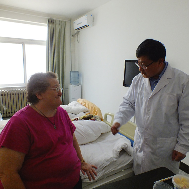
 Nell Smith, a throat cancer patient from Switzerland
 -
 Andress, a 9-year-old boy from the United States
 -
 Anthony, lymphocytic cancer patient from the United States 24
 -
 PAT, rectal cancer patient from the United States
 -
 Mark, a prostate cancer bone metastasis patient from the United States
 -
 Famous American female painter Muriel
Related search
Related search- Cheap gleason 8 prostate cancer treatment near me
- Cheap top cancer hospital near me
- new radiation treatment for lung cancer Hospitals
- treatment early prostate cancer treatment Hospitals
- China primary lung cancer treatment Hospitals
- treatment best prostate cancer treatment centers Hospitals
- Cheap cancer in liver Hospitals
- signs of breast cancer cost
- bladder neck invasion prostate cancer treatment Hospitals
- treatment for brain tumor near me


